Thesis defense: Shijia Wu
| When |
Sep 19, 2025
from 02:00 to 05:00 |
|---|---|
| Where | Salle des thèses |
| Contact Name | Shijia Wu |
| Attendees |
Marie-Stephanie CLERGET-FROIDEVAUX, Professeure Museum National d'Histoire Naturelle de Paris, rapporteure; Beatriz MORTE, Chercheuse, CIBERER, rapporteure; Gérard BENOIT, Chargé de recherche - HDR, Université de Rennes / CNRS, Examinateur; Marie SEMON, Professeur ENS de Lyon, Examinateur; Frédéric FLAMANT, Directeur de recherche, ENS de Lyon / INRAE, Directeur de these; Wenzheng JIANG Professeur, ECNU; Co-directeur de these. |
| Add event to calendar |
|
On September 19th, Shijia Wu of the team of Frédéric Flamant will support her thesis entitled:
"Identification of Thyroid Hormone Receptors Target Genes: In Vitro Analysis and In Vivo Exploration in Mouse Hypothalamus"

Abstract:
Thyroid hormones (THs, including thyroxine and 3,3’,5-triiodo-L-thyronine, or T3, its active metabolite) are essential throughout life. They regulate developmental processes and maintain systemic homeostasis in adulthood, exerting their actions by binding to nuclear receptors (TRs including TRα1 and TRβ1) which are present in virtually all cell types. Typically, TRs form heterodimers with retinoid X receptors (RXRs) and bind DNA to response elements which are widespread in the genome. T3 binding induces chromatin remodeling and activates the transcription of neighboring genes.
The overarching goal of the thesis was to understand the function of THs in the adult mouse hypothalamus, a brain area that is a central regulator of energy balance. The hypothalamus integrates hormone and neural inputs to control temperature, appetite, circadian rhythms, and energy expenditure, all physiological functions, which are sensitive to THs. It also governs the level of circulating THs through the hypothalamus-pituitary-thyroid (HPT) axis. The hypothalamus contains a number of neuronal and glial cell types, and understanding the influence of THs on each of these cell types is a daunting task. To identify TR target genes in hypothalamic cell types, we focused on two criteria: the presence of TR binding sites in gene regulatory sequences and T3-responsive gene expression. As ChIP-seq analysis is currently not feasible in the hypothalamus, we relied on an atlas of TR binding sites identified in a variety of tissues and cell types, a fraction of which being shared across cell types.
The first part of the thesis describes an in vitro analysis of these putative TR response elements. We developed SOSHI-seq (Screening of self-transcribed hormone-inducible elements sequencing), a high-throughput in vitro reporter assay adapted from STARR-seq. In the SOSHI-seq, candidate DNA fragments are placed in the transcribed portion of a plasmid vector. The eventual enhancer activity of a DNA fragment enables it to reinforce its own transcription in a T3-dependent manner. SOSHI-seq is thus a general procedure to test the presence of functional T3-responsive elements in genomic DNA fragments. Screening 2,000 fragments, we identified around 500 T3-activated elements, many containing canonical DR4 motifs (AGGTCANNNNAGGTCA). Luciferase assays confirmed SOSHI-seq findings, and point mutation analyses on specific fragments showed that DR4 motifs were often necessary and sufficient for T3 responsiveness.
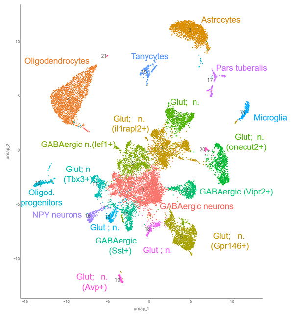
To profile genes in vivo, we treated mice with propylthiouracil to deplete THs. We then treated the mice with THs and performed single-nuclei RNA sequencing (SnRNA-seq) on the mediobasal hypothalamus of mice, combined with bulk RNA-seq in sorted cell types: astrocytes, oligodendrocytes, microglia, GABAergic neurons, and tanycytes. We identified 21 cell clusters, with astrocytes showing the strongest response in SnRNA-seq. Bulk RNA-seq confirmed and extended gene lists.
To examine the physiological roles of T3 signaling, we generated two mouse lines expressing a dominant-negative TRα1 in a cell-type-specific manner, targeting astrocytes and oligodendrocytes, respectively, to block T3 signaling in these populations. Unintended expression of the mutant TRα1 in peripheral tissues, particularly the pancreas, was observed in both lines, precluding an interpretation of the central effects.
In summary, our study provides a multidimensional analysis of T3 signaling in the hypothalamus, combining functional genomics, transcriptomics, and physiological approaches. These findings shed new light on the cell type-specific actions of T3 cells in the brain and highlight the role of the hypothalamus in the regulation of metabolism.