


SBIS, a novel orange fluorescent probe enabling 4D visualisation of living brown algae
The Charrier team has just published a new article entitled:
“SBIS, a new orange fluorescent vital probe for the 4D imaging of brown algal cells” in Journal of Cell Science.
This article is accompanied by a “First Person” feature highlighting the first author, Marie Zilliox.
"I said to myself, “That's it! This is the breakthrough that will truly launch my postdoc project!” I could finally see all the cells in a living embryo."
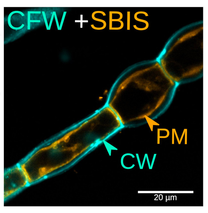
Fig. 1: Labelling of the plasma membrane with the fluorescent probe SBIS (PM, orange) and of the cell wall with the Calcofluor white (CFW, blue) in the filamentous brown alga Ectocarpus. The cell internal pressure has been reduced to better display the plasma membrane.
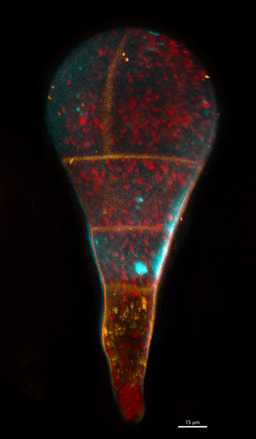
Fig. 2: Embryo of the brown alga Fucus labelled with the orange fluorescent vital probe "SBIS" (red: autofluorescence of the chloroplasts; blue: cellulose present in the cell wall labelled with Calcofluor White).
Read the full article here. The full version of the article is available upon request from Bénédicte Charrier.
Congratulations to the Charrier team for this exciting work!
© Mayeul Collot